



A
Asymmetry
The two halves of the mole not matching each other
Dermatological Treatments
Dermoskopik muayene, bilgisayarlı ben takibi ve gerektiğinde uygun cerrahi yöntemle çıkarma.
Overview
Moles (nevi) are mostly benign growths that may be present from birth or appear later in life. They can be brown, black, skin-coloured or pink, and may be flat or raised.
Most moles are harmless. However, changes that develop over time in some moles, or certain newly appearing pigmented lesions, may require evaluation for melanoma and other skin cancers.
For this reason, regular dermatological examination and, when needed, dermoscopic mole monitoring are important for people with many moles, atypical moles or a family history of melanoma.
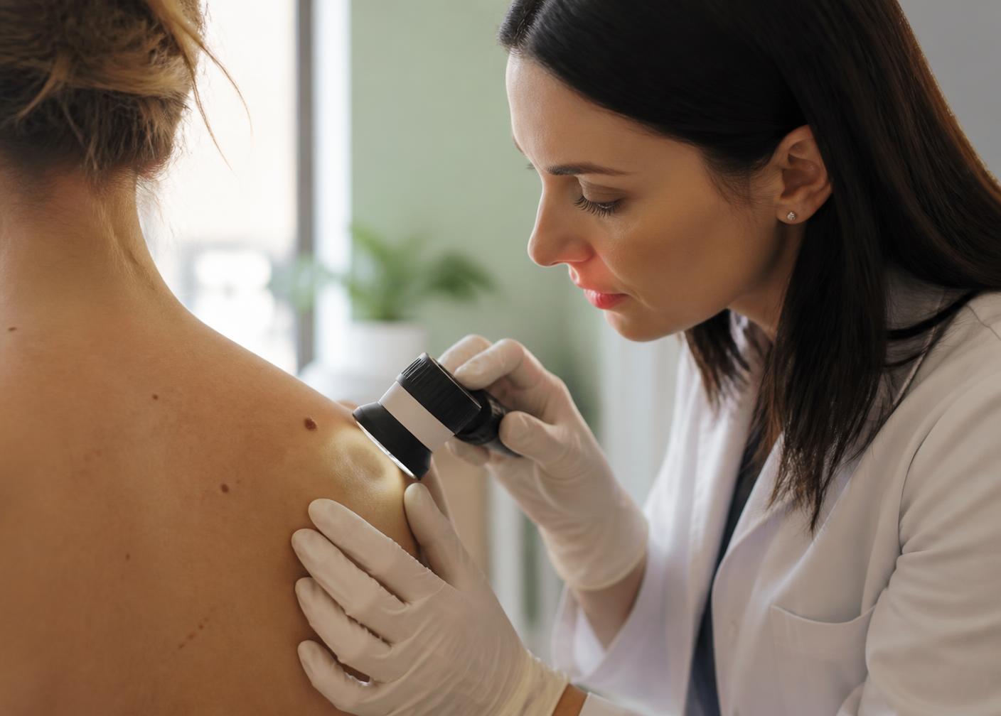
During a mole examination, lesions on the skin are first assessed dermatologically. Moles that require closer inspection are magnified and examined with a special device called a dermatoscope.
Dermoscopy helps evaluate colour and structural features that cannot be seen with the naked eye.
Computerised dermoscopic follow-up can be used for people with numerous or atypical moles. Images of the moles are recorded and compared with previous images at subsequent visits.
This makes it possible to follow small changes in moles over time more objectively.
Appointment
Your moles are assessed with clinical examination and dermoscopy; the follow-up interval is set individually.
Appointment · +90 533 551 41 62Follow-up
Particularly;
moles should be evaluated by a dermatologist.
Self-Monitoring
The ABCDE rule can be a guide for personal mole monitoring:
A
The two halves of the mole not matching each other
B
Irregular borders
C
More than one colour or uneven colour distribution
D
Especially enlarging lesions
E
Change in the size, shape, colour or structure of the mole over time
However, the ABCDE criteria alone cannot determine whether a mole is benign or malignant. This is why dermoscopic assessment is important.
Digital Monitoring
Bilgisayarlı dermoskopi, benlerin yüksek çözünürlüklü dermoskopik görüntülerinin dijital ortamda kaydedilmesine ve zaman içerisinde karşılaştırılmasına olanak sağlayan bir takip yöntemidir.
Particularly;
Not every mole needs to be removed. Regular monitoring of non-suspicious moles is usually sufficient.
Mole Removal
01
The method used for mole removal is determined by the type, size and location of the mole and the reason for removal.
For moles suspicious for melanoma or another skin cancer, the lesion must be removed with an appropriate surgical technique and sent for pathological examination.
The procedure is performed under local anaesthesia. After the mole is removed, the area is closed with sutures when necessary.
02
Histopathological examination is important for pigmented lesions that are suspicious for skin cancer or require a confirmed diagnosis. A suspicious mole should never be removed by cosmetic methods alone; an accurate diagnosis must come first.
03
Some moles, although benign, may bother a person because of their location on the face or body, or may be constantly irritated by clothing, jewellery or shaving.
For suitable lesions with benign features on dermatological and dermoscopic assessment, surgical excision, shave excision or other methods may be used depending on the lesion. The most appropriate method is decided according to the structure and location of the mole.
Safety
No. Removing a mole appropriately does not cause cancer.
What matters is that a suspicious mole is properly evaluated before the procedure and sent for pathological examination when needed.
Recovery
Any procedure that breaks skin integrity carries some possibility of scarring.
The visibility of the scar depends on;
varies accordingly.
For a mole that must be removed surgically, the priority is an accurate diagnosis and appropriate excision of the lesion.
Frequently Asked Questions
The aim is not to remove every mole, but to notice suspicious changes early.
No. Most moles are benign and do not need to be removed. Suspicious moles are evaluated; those that require it are removed, while others can be monitored.
The frequency depends on the number of moles and individual risk factors. For people with numerous or atypical moles, follow-up intervals are set after a dermatological examination.
No. New moles can appear throughout life. However, lesions that newly develop and grow in adulthood, or that look different from other moles, should be evaluated.
Before applying a laser to a pigmented lesion, the diagnosis must be certain. Laser is not preferred for lesions suspicious for melanoma; the lesion is removed appropriately and sent for pathological examination.
Since the procedure is performed under local anaesthesia, pain is not expected during it. Short-term tenderness may occur afterwards.
Assessment
The goal of mole monitoring is not to remove all moles, but to detect suspicious changes at an early stage.
Through clinical examination, dermoscopy and, where needed, computerised dermoscopic follow-up, moles can be compared over time.
For lesions considered suspicious, the aim is a definitive diagnosis through appropriate surgical removal and pathological examination.
Dermatological Treatments
A plan that stops active acne and gradually repairs scars.
View →A control plan for persistent redness, visible capillaries and sensitivity.
View →Skin health, barrier repair, and revitalization protocols.
View →Personalized protocol for melasma, sun spots, and post-inflammatory marks.
View →Diagnosis and treatment for fungal infection, ingrown nails and colour changes.
View →